Babyecho for Dummies
:max_bytes(150000):strip_icc()/191127-ultrasound-trimester-pink-2000-fd089add04f8444e9d7a403933d1994f.jpg)
For most females, ultrasound reveals that the child is expanding usually. If your ultrasound is regular, simply be certain to keep mosting likely to your prenatal appointments. Occasionally, ultrasound might reveal that you and your baby need special treatment. For instance, if the ultrasound reveals your infant has spina bifida, he may be treated in the womb prior to birth.
A c-section is surgical treatment in which your baby is birthed through a cut that your medical professional makes in your stomach and uterus. Regardless of what an ultrasound reveals, talk with your supplier regarding the very best treatment for you and your infant - baby heartbeat doppler. Last examined: October, 2019
Throughout this check, they will examine the baby is expanding in the ideal place, whether there is greater than one baby and they will certainly additionally check your infant's development until now. This testing is available in between 10 14 weeks of pregnancy and is used to analyze the possibilities of your child being birthed with several of these conditions.
Babyecho Things To Know Before You Buy
It entails a combined test of an ultrasound check and a blood examination. During the scan, the sonographer will determine the fluid at the rear of the baby's neck to figure out 'nuchal clarity' - https://www.indiegogo.com/individuals/37855747. They will after that compute the opportunity of your child having Down's, Edwards' or Patau's syndrome utilizing your age, the blood test and scan outcomes
Throughout this scan, the sonographer look for structural and developmental problems in the child. During this check consultation, you might be offered testings for HIV, syphilis and hepatitis B by a specialist midwife. In many cases, a third-trimester check is suggested by your midwife adhering to the outcomes of previous tests, previous complications or existing medical problems.
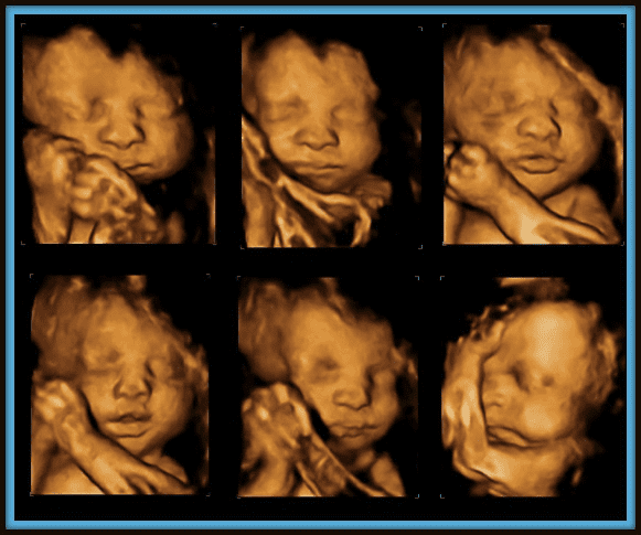
The traditional 2D ultrasound creates flat and described photos which can be made use of to see your child's interior organs and help identify any type of internal concerns. These black and white pictures aid the sonographer establish the child's gestation, development, heartbeat, development and dimension. Some expectant mothers choose to have a 3D ultrasound check due to the fact that they reveal more of a real-life picture of the baby.
Excitement About Babyecho
3D ultrasound scans show still images of your child's outside body as opposed to their withins, so you can see the form of the infant's face attributes. 4D ultrasound scans are similar to 3D scans but they show a relocating video clip instead of still pictures. This records highlights and darkness better, consequently developing a clearer photo of the child's face and activities.

or (8-11 weeks) (11-14 weeks) (14-18 weeks) (19-23 weeks) or (24-42 weeks) Suggested at Optional -, extra often in some problems This scan is done to and to identify an (EDD). A is discovered during this scan. Many parents go with this scan for. Is important prior to the blood test called as (NIPT) to compute the.
Some Known Details About Babyecho
Sometimes a may be called for to obtain and a more clear photo. This is normally done and occasionally a may be needed (doppler ultrasound). Confirm that the child's heart is existing; To extra accurately.
Please see below. These scans may be done, however some of the and for this reason, a is required to This check is done generally at.
The Buzz on Babyecho

In addition, the can be by by an. and is kept track of by these scans. of, andare done to reach an. around the child is measured. and child's are checked. () The method nearer the serves to. Periodically, an which was previously might be.
The Best Guide To Babyecho
If, these scans may be to. (of the baby) can also be carried out. This includes, along with; This consists of, along with (14-20 weeks).
A check is essential prior to this examination is done. If you're looking for, organize an examination with Dr Sankaran by means of her. Obstetrics & gynaecology in London.
The 7-Second Trick For Babyecho
The test can give useful details, aiding women and their health-care providers handle and care for the pregnancy and the unborn child.
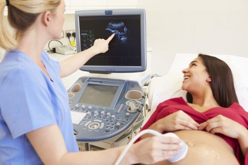
A transducer is placed into the vagina and rests against the back of the vaginal canal to produce a photo. A transvaginal ultrasound creates a sharper photo and is commonly used in very early maternity. Ultrasound devices have to do with the size of a grocery store cart. A television screen for checking out the photos is connected to the machine (https://pagespeed.web.dev/analysis/https-babyfetaldoppler-com/wlbdlhbwfi?form_factor=mobile).
Comments on “Facts About Babyecho Revealed”